Fortunately, technology like flow imaging microscopy (FIM) can reduce staff time and variability. FIM combines the optical benefits of microscopy with the speed of particle analysis to automatically detect, image, enumerate, and measure particles in flow. FIM instruments like FlowCam enable users to accelerate a variety of applications in ways that are not practical through microscopy alone.
Shellfish Reproduction
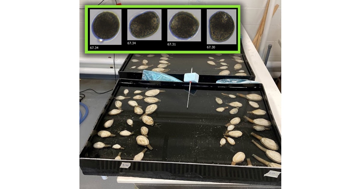
Egg density and viability are critical to the success of shellfish spawning and breeding. Oysters, mussels, and clams can produce millions of eggs per spawning event. FIM is ideally suited to image, count, measure, and characterize these shellfish eggs both pre- and post-fertilization.
Egg size is a critical data point, and egg shape can be used to detect abnormalities that indicate underdevelopment or poor conditioning. Additionally, images differentiating fertilized from unfertilized eggs based on the presence of polar bodies can help maximize the data from a single sample. FlowCam®, a flow imaging microscope, is used by labs like the Downeast Institute (Maine, US) to accelerate particle counting and measurement during each spawn. With FlowCam fertilization, success rate, egg growth, and abnormalities can be quickly and reliably evaluated.
Tessa Houston, a Research Assistant at the Downeast Institute, comments on using FlowCam, “The post-spawning analysis was so much easier with FlowCam. We used to spend hours looking at the microscope, analyzing upwards of 30 samples (10–15 minutes per sample). With FlowCam, this process took us four minutes per sample.”
Shellfish Larvae
Tracking the growth and condition of shellfish larvae is a core function of any hatchery or shellfish research program, but counting and measuring larvae on a microscope is time consuming and prone to variability between operators: even a minor adjustment in the fine focus between operators can produce a 5+ μm difference in size.
Rapidly count and measure larvae at several developmental stages, assess their viability, and monitor larval transport. FlowCam’s flow imaging software automatically generates data on length, width, and area in a consistent and reproducible way while simultaneously determining larval concentration. Additionally, FlowCam’s color camera may be used to differentiate live larvae from dead larvae using a Neutral Red stain. Certain FlowCam models can even be used to efficiently analyze high-density samples for mark-recapture studies to track calcein-stained larval dispersal in the field; understanding larval transport is critical for shellfish management and restoration.
FlowCam is used by labs like NOAA’s Milford Lab to automatically enumerate, measure, and assess shellfish larvae, producing image data faster than was possible with manual microscopy.

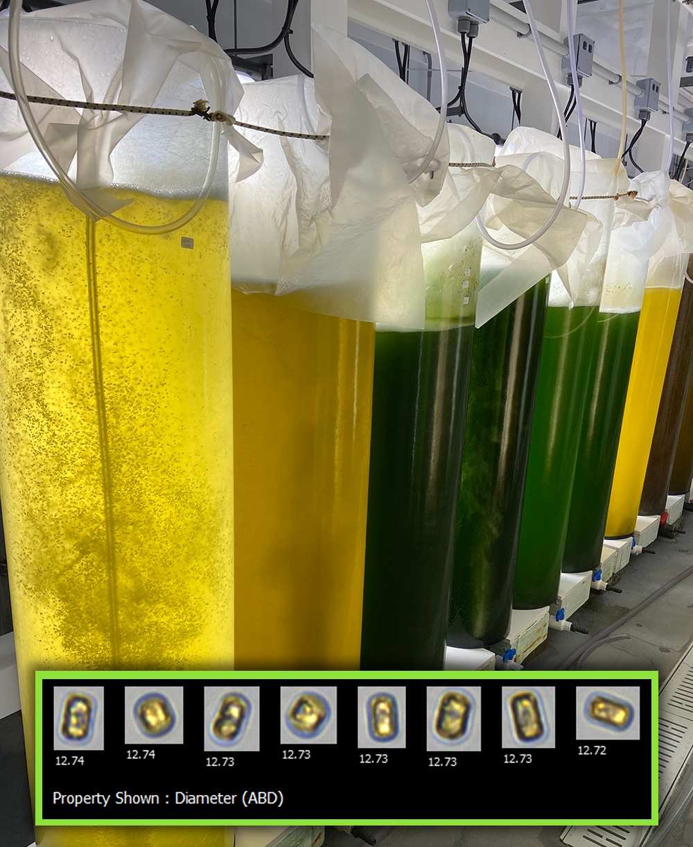
Algae growth tanks in Downeast Institute’s mass culture room. Inset: Thalassiosira weissfloggi cells, imaged by FlowCam. (Image credit: Yokogawa Fluid Imaging Technologies)
Phytoplankton Analysis
One of flow imaging microscopy’s core applications is the analysis of phytoplankton communities and their size distribution. This application extends to shellfish aquaculture, where FIM can be used not only for evaluating shellfish eggs and larvae but also the phytoplankton they feed on throughout their life cycle—both in the hatchery and in the field.
Quality monitoring of algae production requires information on the quantity of algae and the health of the cultures. FIM can provide both types of data simultaneously and with a high level of reproducibility, allowing both algae cultivated in the hatchery and marine phytoplankton in shellfish growout locations to be analyzed quickly, easily, and economically.
Predators
FlowCam’s magnification options enable users to capture top-down pressure from environmental threats to adult shellfish—targeting not only small particles like eggs, larvae, and phytoplankton but larger organisms, too. Predators like invasive crabs can threaten adult shellfish during growout. These crabs start their life cycle as eggs and larvae that can also be detected and measured using flow imaging microscopy.
Combined with genetic techniques, FlowCam offers a powerful tool to visualize and count possible predators in their larval forms to better characterize when and where they spawn. FIM has been utilized by Ph.D. students like Kelsey Meyer at the University of New Hampshire to help better understand and quantify this threat to the shellfish aquaculture industry in key growing areas thanks to FlowCam’s ability to target oyster larvae and crab larvae with a single instrument.
Kelsey Meyer, a Ph.D. student at the University of New Hampshire, commented: “FlowCam offered us a reliable and objective means of counting cells and particles in our water samples. It was great to be able to pinpoint both oyster and crab larvae. We created libraries and set up a classification scheme to automatically identify and count oyster and crab larvae, and having FlowCam provide this data objectively was fantastic.”
In Action
FlowCam combines the optical elements of a microscope with the speed of a particle counter. Some FIM instruments even utilize principles of flow cytometry by incorporating a laser to trigger image capture based on pigments like chlorophyll.
FlowCam consists of a light microscopy optics system paired with a flow cell and digital camera. A syringe pump pulls a water sample through the flow cell, and images of particles passing through are automatically captured, counted, measured, and saved.
FlowCam’s built-in software, VisualSpreadsheet®, is a powerful and flexible program for both data acquisition and image analysis. Some of the morphological properties determined by VisualSpreadsheet include diameter, area, aspect ratio, circularity, color, transparency, and intensity. The user can filter and sort particles according to their properties and display results in interactive scattergrams or histograms.
Sophisticated pattern recognition allows researchers to immediately find and display organisms with similar properties or create image libraries and classification templates that can be applied to new samples to help determine concentrations of specific populations.
FlowCam’s speed, optics, and image recognition capabilities are among the many reasons shellfish hatchery managers are turning to FIM to help make the process of counting and evaluating shellfish eggs and larvae faster and more reproducible.
To find out more about FlowCam, visit: www.flowcam.com
This feature appeared in Environment, Coastal & Offshore (ECO) Magazine’s 2024 Summer edition Fisheries & Aquaculture, to read more access the magazine here.

