For more than 400 million years, sharks have evolved a unique cartilage-based skeleton that provides the strength and flexibility needed to endure millions of powerful movements. While scientists understand how shark muscles drive different swimming styles, far less is known about how the internal structure of their spines helps them achieve remarkable speed, agility, and endurance.
Now, Florida Atlantic University researchers and collaborators from NOAA Fisheries are taking a closer look inside the shark spine to uncover how its hidden design enables these ancient predators to move with such power, precision, and stamina.
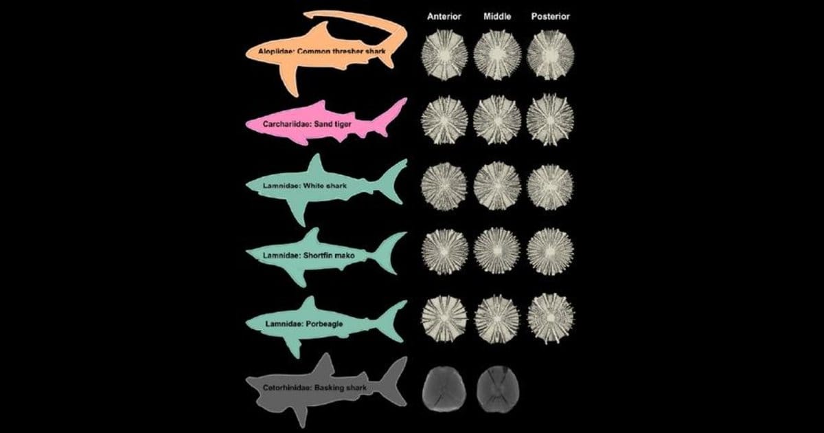
The new study, published in the Journal of Anatomy, reveals that the internal architecture of shark vertebrae is remarkably specialized, with each species evolving a vertebral column uniquely suited to the way it moves through the water. By examining the intricate mineralized structures inside the cartilaginous vertebrae of six species—the great white, shortfin mako, porbeagle, common thresher, sand tiger, and basking shark—researchers discovered that the spine is far more than a flexible support column. It’s a finely engineered biomechanical system designed to maximize strength, flexibility, and swimming efficiency.
To uncover these hidden adaptations, the research team analyzed vertebrae collected from different regions of each shark’s spine. Using high-resolution micro-computed tomography (micro-CT), they created detailed 3D images that allowed them to look inside the vertebrae without damaging them.
The researchers measured the size and shape of each vertebra and mapped tiny mineralized plates and branching structures, comparing how these features changed from the front of the body to the tail and among species with different swimming speeds.
“High-resolution micro-CT imaging gave us a window into the hidden world inside shark vertebrae, allowing us to see structures that have evolved over hundreds of millions of years to support these incredible swimmers,” said Jamie Knaub, first author, research specialist at FAU Laboratory Schools and a Ph.D. candidate in the FAU Department of Biology within the Charles E. Schmidt College of Science.
Knaub scanned the vertebrae investigated in the study at the Berlin Family Bioimaging Lab at FAU Lab Schools Marcus Research and Innovation Center.
“By examining species with varied swimming strategies, we found that the shark spine is not a one-size-fits-all design,” said Knaub. “Instead, its internal architecture reflects the unique demands of each species’ movement.”
The findings showed that the middle of the spine, where there may be a ‘hinge’ in the wave produced during swimming, consistently contained the largest vertebrae. But the biggest surprise came from what was happening inside them.
Fast-swimming species such as the great white, shortfin mako, and porbeagle had vertebrae with internal mineralized structures arranged to create a stiffer vertebral column that efficiently transfers energy to the tail, helping propel these sharks through the water at high speeds. In contrast, the shape and structure of sand tiger shark vertebrae likely provide greater flexibility, supporting slower, more maneuverable swimming through complex underwater environments.
Common thresher sharks, famous for using their exceptionally long tails to stun prey, displayed the largest quantity of mineralized plates and branching structures that appear adapted to withstand the powerful side-to-side and overhead tail strikes unique to their hunting strategy. Meanwhile, the basking shark—the world’s second-largest fish and a slow-moving filter feeder—had vertebrae with dramatically reduced mineralization, reflecting the very different mechanical demands of cruising through the ocean while feeding.
The researchers also found that the arrangement of these mineralized structures changes along the length of the spine, particularly near the tail where swimming forces are greatest. In fast-swimming sharks, the rear vertebrae contained more mineralized plates, making this region stiffer and more efficient at transferring energy during every tail beat.
“Nature has spent hundreds of millions of years refining these designs,” said Marianne E. Porter, Ph.D., senior author and FAU Department of Biological Sciences professor. “What we see is an elegant example of biomechanics in action, where each species has evolved a vertebral column precisely tuned to the way it swims. The shark spine isn’t simply flexible—it’s optimized to balance strength, stiffness, and motion in ways that maximize performance.”
The study also revealed that closely related species, such as great white and shortfin mako sharks, share remarkably similar vertebral designs, while more distantly related sharks have evolved distinctly different internal architectures.
“This work would not have been possible without advanced micro-CT technology, which lets us visualize complex 3D structures that were previously inaccessible,” said Tricia L. Meredith, Ph.D., co-author and Director of Research for FAU’s on-site lab schools, A.D. Henderson University School and FAU High School, and an assistant research professor in FAU’s College of Education. “These imaging capabilities allow us to move beyond simply describing anatomy to understanding how internal structures function mechanically, opening the door to new discoveries in comparative biology and biomimetic design.”
Beyond advancing knowledge of shark biology, the research could have broader implications for engineering. Understanding how sharks combine lightweight cartilage with strategically placed mineralized architecture may inspire new materials and technologies that require both flexibility and strength, from robotics to biomedical devices.
Study co-authors are FAU undergraduate students of biological sciences Madisan Biordi and Emma Pawlik; Michelle Passerotti, Ph.D., Apex Predators Program leader, NOAA Fisheries; and Lisa J. Natanson, Ph.D., researcher at NOAA Fisheries.